Oral, Dental, and Maxillofacial Radiology is a subspecialty field of dentistry that aims to make accurate diagnoses of diseases and plan appropriate treatments.
Oral Diagnosis and Radiology is the first specialized field patients encounter when they visit a dentist with complaints about oral, dental, or soft tissue issues. Managing the process from meeting the patient to treatment planning with accurate information is important for breaking the "dentist" phobia and gaining the trust of patients in every way.
Determining the medical issues in the patient's past and present history, conducting a detailed intraoral and extraoral examination, interpreting periapical-panoramic-tomographic X-ray findings, making accurate differential diagnoses of diseases, and planning the appropriate treatment based on all this data are the skills of this subspecialty field.
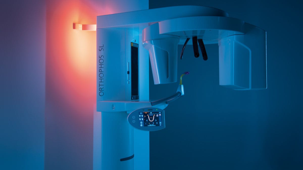
ORTHOPHOS SL 2D
We provide services with X-ray devices that facilitate advanced diagnostic options for endodontics, periodontics, implantology, orthodontics, surgery, and other specialized departments.
Thanks to Sharp Layer Technology (SL), high-resolution panoramic images are not only obtained in the sharp layer, but also enable analysis in the lingual/buccal plane in special cases.
This imaging device has a viewing area starting at 5cm x 5cm and extending up to 11cm x 10cm for upper respiratory tract analysis.
ORTHOPHOS SL 2D, with DCS (Direct Conversion Sensor) sensor and SL technology, provides images that satisfy even dentists with high demands for panoramic imaging. With its 3-point head immobilization, balanced arms, and patented occlusal bite rod, it guarantees unique resolution in every image. Together with the pioneering SIDEXIS 4 software, it offers a unique variety of innovative solutions for clinical workflow.
Our hospital's ORTHOPHOS SL device, which perfectly fits the pleasant atmosphere, provides relaxing ambient lighting with more than 30 colour options, which also has a special feature for patients with claustrophobia, enabling our patients to have a more comfortable, smooth, and fast imaging.
Most of the patients who visit our hospital are hesitant to visit the radiology department due to the amount of radiation from dental X-rays. However, there are some differences between our hospital's ORTHOPHOS SL device and the X-ray devices used in other hospital institutions or clinics:
- By taking quick shots within 15 seconds, the radiation dose the patient receives is cut in half.
- With its sharp X-ray feature, it automatically gathers and combines sharp layers in films according to the patient's personal anatomical structure, providing HD images to our dentists, making it easier for them to make more accurate diagnoses.
- Thanks to the Sharp Layer Technology (SL) technology, high-resolution panoramic images are not only obtained in the sharp layer, but also enable interactive analysis in the lingual/buccal plane in special cases.
- Our X-ray device with HD mode capability allows us to see the thin root canals in the patient's jaw structure with high-quality images even in panoramic films.
- Did you know that the radiation dose from our SIRONA ORTHOPHOS SL 2D/3D X-ray device is less than the radiation dose from X-ray shots taken with other devices used in hospitals and clinics, as well as the radiation dose you receive in your daily life?
In the table below, the radiation doses mentioned are compared with the radiation dose given by our ORTHOPHOS SL device. As a result, it is concluded that each device does not give the same radiation dose, and that the devices used change the amount of radiation received.